The short version
- Histotripsy aims short pulses of focused ultrasound through the skin to make bubbles inside a tumor, and the bubbles tear the tissue apart without heat or a needle.
- In the 44-patient trial behind its FDA clearance, the treatment destroyed the planned target 95% of the time, and 7% of patients had a serious complication.
- At one year, scan readers judged 63% of tumors controlled on the first read and 90% on a second read, partly because blood vessels the treatment spares can look like tumor.
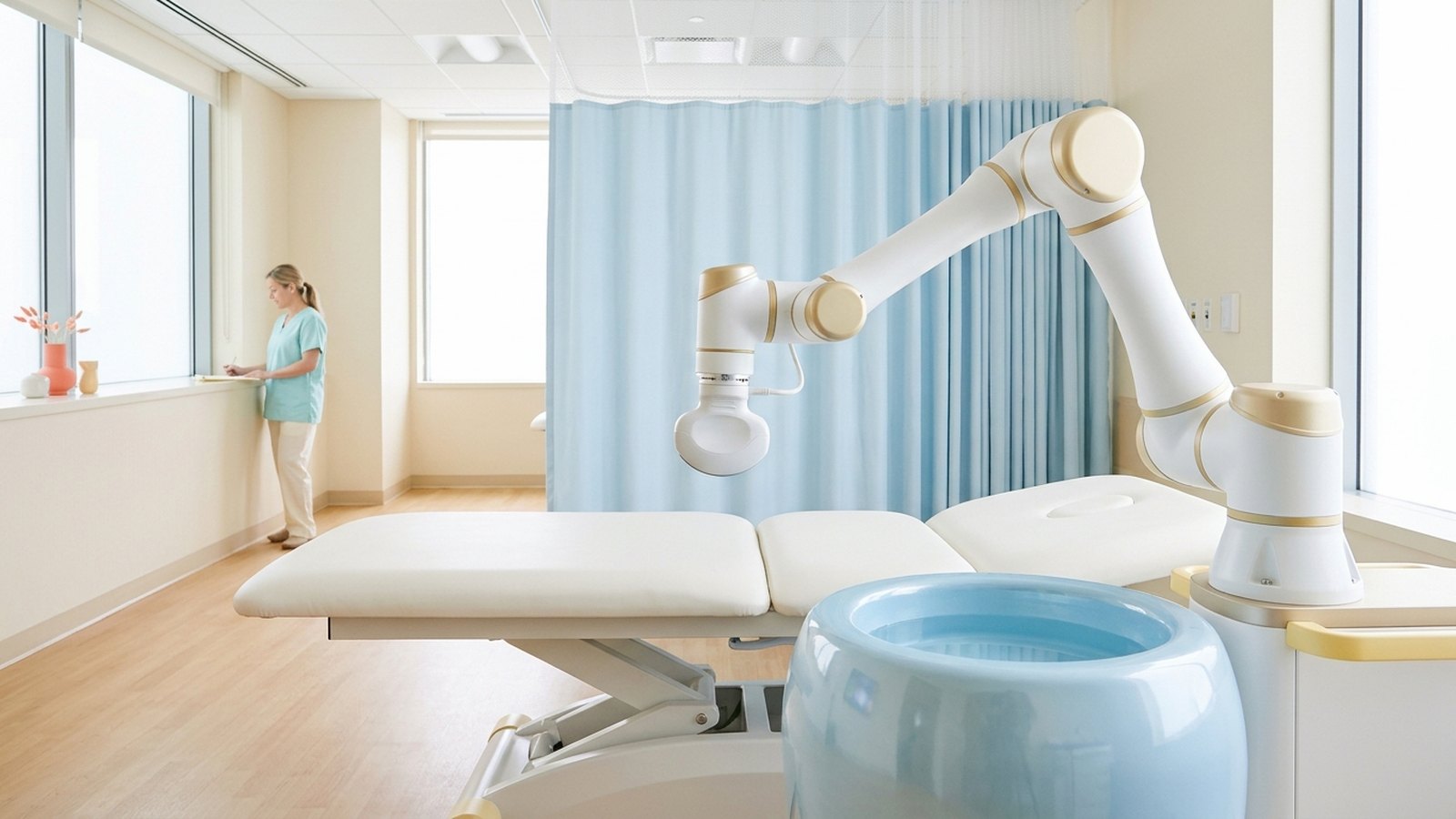
A robotic arm holds an ultrasound head over a patient’s abdomen, guided by an ultrasound image of the liver. Nobody cuts anything. The machine fires pulses of sound that last less than 20 millionths of a second each, and a few centimeters under the skin, a tumor about 1.5 centimeters across is broken down into liquid.
That is histotripsy. The FDA cleared the first system, HistoSonics’ Edison, for liver tumors in October 2023. This year it was approved in Taiwan in May, in Europe in July and in Singapore last week, which we covered on Wednesday. The company has asked the FDA to add kidney tumors.
Approvals are arriving quickly. The evidence behind them comes mostly from one company-sponsored trial programme, so it is worth reading that trial closely.
What the sound actually does
Many ultrasound treatments work by heat: focus enough sound on one spot and the tissue warms until it is destroyed. The trial paper describes histotripsy as nonthermal. Each pulse is very short and very intense. At the focal point, the negative half of the pressure wave pulls so hard that gas in the tissue bursts into a cloud of tiny bubbles, which then collapse. This is called cavitation, and pulse after pulse, it breaks the cells down mechanically into liquid.
The pulses run at 700 kHz and last under 20 microseconds. Each focal point destroys a patch about 3 × 3 × 6 millimeters, and the robotic arm steps that point through the planned volume while an ultrasound image guides it. The system reaches 14 to 16 centimeters deep.
The proposed advantage is what it leaves behind. Animal work cited by the trial team suggests tougher, collagen-rich structures such as blood vessels and bile ducts survive pulses that destroy softer tumor tissue. That detail turns out to matter a great deal when it comes to reading the results.
The trial behind the approval
The evidence for the FDA decision is #HOPE4LIVER, two parallel trials in the United States and in Europe and England, published in Radiology in September 2024. The work was sponsored by HistoSonics, and several authors have financial ties to the company.
Between January 2021 and July 2022, 14 hospitals treated 44 adults. Eighteen had hepatocellular carcinoma, the most common primary liver cancer, and 26 had cancer that had spread to the liver from elsewhere, mostly the colon, pancreas or breast. Each could have up to three tumors smaller than 3 centimeters. The average largest tumor was 1.5 centimeters across.
The trial had two pass-fail goals, agreed in advance with the FDA. The first was technical success: did the treatment destroy the planned target, as seen on a scan within 36 hours? The goal was over 70%. The result was 42 of 44 tumors, or 95%.
The second goal was safety: fewer than 25% of patients with a major complication within 30 days. Three of 44 had one, or 7%. One patient with a biliary stent developed sepsis and was treated with antibiotics. One had chest pain from the lining of the lung and spent two days in the hospital. And one patient, whose liver was heavily loaded with metastatic breast cancer, went into liver failure on day 12 and died on day 37.
Both goals were met. The authors also report that a core laboratory found damage outside the target in 6 patients, or 14%.
The number that depends on who is looking
Destroying a tumor on the day is the first test. The one that matters is whether it stays gone. In 2025 the same team published one-year results in Annals of Surgery, now covering 47 patients and 52 tumors.
An independent core laboratory read every follow-up scan as it came in, using the rules developed for heat-based ablation, in which a bright nodule at the edge of a treated zone counts as tumor coming back. By that primary reading, 63.4% of tumors were controlled at one year.
Then the same laboratory re-read the scans after every patient had finished a year, this time looking at each patient’s whole series at once. By that second, post hoc reading, local control was 90%.
The authors explain the gap. Histotripsy can leave blood vessels intact inside the treated zone, and on a scan those surviving vessels can look like the bright nodules the old rules treat as tumor. None of the trial’s doctors had used histotripsy on a liver before, so the second read was meant to allow for that learning curve. The two readings agreed between 71% and 84% of the time.
That explanation is plausible, and it fits what the animal work predicts. It is also, by definition, a judgment made after the fact. The Focused Ultrasound Foundation’s summary of the paper reported only the 90%. Only 30 of the 52 tumors were followed to one year, and 23 had scans clear enough for the full analysis.
One-year survival was 73.3% for patients with primary liver cancer and 48.6% for those with spread cancer. The authors call both comparable to other treatments for similar stages of disease. The trial had no comparison group to check that against, and later treatments patients received were not consistently tracked. Ninety-five percent of participants were white.
The wider evidence
A meta-analysis in EClinicalMedicine this spring, led by Chase Wehrle at the Cleveland Clinic, pooled 10 studies and 553 patients. Technical success was 94.1%, and serious complications, in the four studies that reported them, ran at 7.0%, with a wide margin of uncertainty. The authors noted that the studies measure tumor control in different ways and follow patients only briefly.
So the honest summary is this. Histotripsy reliably destroys small liver tumors on the day, with serious complications in about 1 patient in 14. Whether it controls them as well as heat-based ablation or surgery over years is not known, because no trial has compared them directly. The best one-year figure available depends on a re-read of the scans that the trial’s own authors describe as post hoc.
None of that makes it a bad tool. A noninvasive treatment that spares vessels and bile ducts could matter most for tumors next to them, where heat is hard to use safely. It just means the case is still being made.
What to watch
The #HOPE4LIVER patients will be followed for five years, and the authors say future studies will add biopsies after treatment, which would settle what those bright spots on the scans really are.
Kidney is next. The #HOPE4KIDNEY trial enrolled 67 patients with biopsy-proven kidney tumors of 3 centimeters or less and measured safety at 30 days and tumor destruction at 90 days. It has been submitted to the FDA, but no results have been published. William Huang, chief of urology at NYU Langone’s Tisch Hospital, put the goal plainly in an interview with Urology Times this week: “We want to provide oncologic control and yet preserve kidney function to the best that we can do.”
The question that will decide histotripsy’s place is the one no trial has yet asked: patient for patient, how does it compare with the treatments it hopes to replace?